Test, Treatment & Technology
Tests, treatment & Technology
Tests
Optical Coherence Tomography (OCT)
OCT is short for Optical Coherence Tomography, a modern imaging technique, which shows structures inside the eye that can change due to eye disease.
In an OCT exam, a light beam scans across the single retinal layers. The beam scans across the back of the eye, and the reflected light is translated into a detailed image of the structures within the retina.
OCT has become invaluable in advanced eye care because it allows your doctor to see tiny changes in the eye, which would otherwise be difficult to detect.
Heidelberg OCT

OCT of macula

Copyright notice: This image was originally published in the Retina Image Bank® website
Author: Jennifer Carstens, Photographer:Jing Zhang
Title: CSNB-OCT-OS, Year: 2021, Image Number:81995
© The American Society of Retina Specialists.

OCT of Macula
Copyright notice: This image was originally published in the Retina Image Bank® website
Author: Jennifer Carstens, Photographer:Jing Zhang
Title: CSNB-OCT-OS, Year: 2021, Image Number:81995
© The American Society of Retina Specialists.

Eiden OCT
Fundus Photography
- Early Detection of Eye Conditions
- Baseline Documentation
- Monitoring Chronic Conditions
- Customized Treatment Plans
- Patient Education
- Documentation for Referrals
- Research and Education
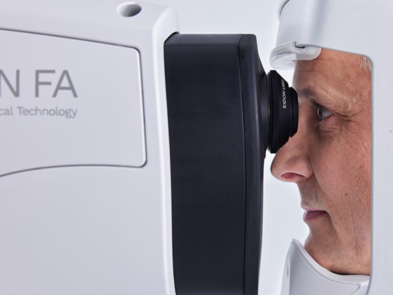
Fundus Camera
Color fundus image
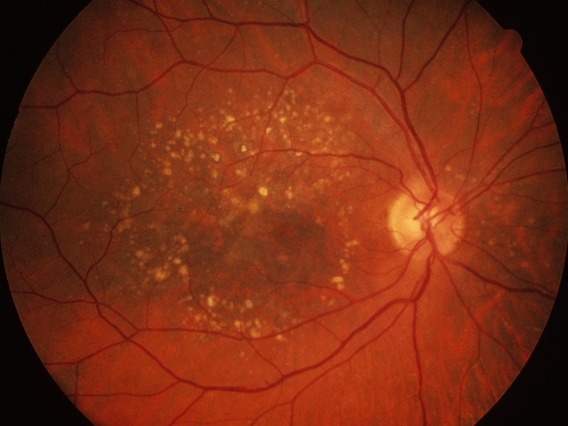
Copyright notice: This image was originally published in the Retina Image Bank® website ©
Author: Henry Kaplan, MD Photographer: Henry Kaplan, MD
Title: Dry AMD, Year: 2014, Image Number: 18286
©The American Society of Retina Specialists.
Fundus Camera
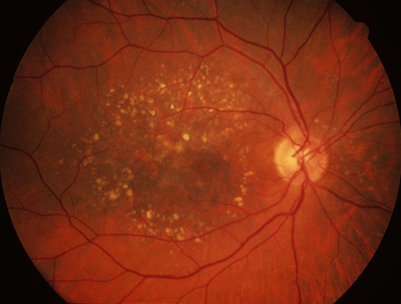
Macular Degeneration
Copyright notice: This image was originally published in the Retina Image Bank® website ©
Author: Henry Kaplan, MD Photographer: Henry Kaplan, MD
Title: Dry AMD, Year: 2014, Image Number: 18286
©The American Society of Retina Specialists
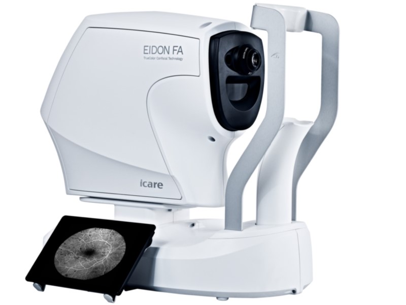
Fluorescein Angiography
Fluorescein angiography is a diagnostic procedure used to assess the blood vessels in the retina and choroid (the vascular layer beneath the retina) of the eye. It involves the use of a fluorescent dye called fluorescein, which is injected into a vein, usually in the arm. The dye travels through the bloodstream and illuminates the blood vessels in the eye when it reaches the retina. This procedure can be used for the following reasons:
- Diagnosing Retinal Diseases
- Detecting Abnormal Blood Vessel Growth
- Monitoring Disease Progression
- Guiding Treatment Decisions
- Assessing Retinal Blood Flow
- Research and Education
Fundus Camera

fluorescein angiography
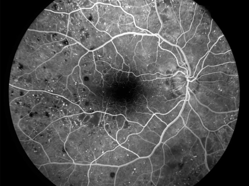

Fundus Camera

Fluorescein Angiography
Visual Field Testing
Visual field testing, also known as perimetry, is a diagnostic procedure used to assess a person’s entire field of vision, including their central and peripheral vision. At Texas Macula & Retina, we use cutting edge technology that allows the use of virtual reality to test your visual field. This allows you to sit in the comfort of your own chair and relax while the test is occurring. Visual field testing can be used for the follow functions:
- Detecting Eye Diseases
- Glaucoma Management
- Evaluating Neurological Disorders
- Monitoring Progression
- Assessing Functional Vision
- Driver’s License Renewal
Ocular Ultrasound
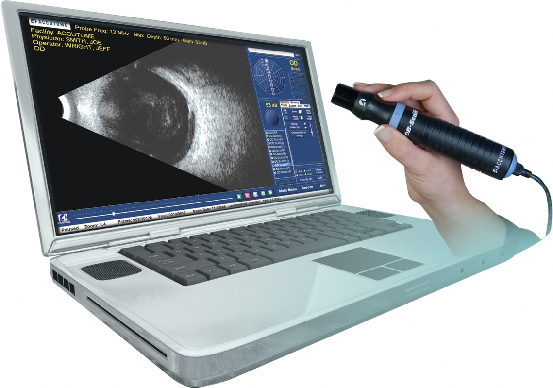
Ocular ultrasound or simply “B-scan”, is a diagnostic imaging technique used to visualize the structures inside the eye. It is especially useful in situations where a direct view of the eye’s interior is obstructed or when assessing structures that are not easily visible through traditional ophthalmoscopy. It can be used for the following purposes:
- Non-Invasive Visualization
- Diagnostic Tool
- Assessment of Eye Trauma
- Monitoring Eye Diseases
- Evaluation of Intraocular Tumors
TREATMENTS
Injection
Intraocular injections, also known as intravitreal injections, are a medical procedure in which medication is injected directly into the vitreous cavity, the gel-like substance that fills the middle of the eye. These injections are important for several reasons in the field of ophthalmology:
- Targeted Delivery of Medication: Intraocular injections allow for the precise and targeted delivery of medications to the interior of the eye. This is particularly important for treating conditions that affect the posterior segment of the eye, such as the retina, macula, and vitreous, where systemic medications may not reach therapeutic levels.
- Treatment of Retinal Diseases: Intraocular injections are commonly used to treat various retinal diseases, including age-related macular degeneration (AMD), diabetic retinopathy, retinal vein occlusions, and macular edema. Medications injected into the vitreous can directly address these conditions and help reduce vision-threatening complications.
- Management of Eye Inflammation: Intraocular injections can be used to deliver anti-inflammatory medications, such as corticosteroids, to manage conditions like uveitis or macular inflammation. These injections can help reduce inflammation within the eye and alleviate symptoms.
- Preservation of Vision: In many cases, intraocular injections are used to prevent or slow the progression of sight-threatening eye diseases. By delivering medication directly to the source of the problem, these injections can help preserve and improve a patient’s vision.
- Alternative to Surgery: In some instances, intraocular injections may serve as an alternative to surgical interventions. For example, injections are often used before considering laser therapy or vitrectomy (surgery) for certain retinal conditions.
- Minimized Systemic Side Effects: Intraocular injections minimize systemic side effects because the medication is delivered directly to the eye, reducing the need for large systemic doses of medications.
Intraocular injections are typically administered by retina specialists and are considered safe when performed by trained professionals. The frequency and duration of injections depend on the specific condition being treated and the patient’s response to therapy. Overall, intraocular injections have revolutionized the treatment of many sight-threatening eye diseases, offering the potential for improved visual outcomes and enhanced patient quality of life.
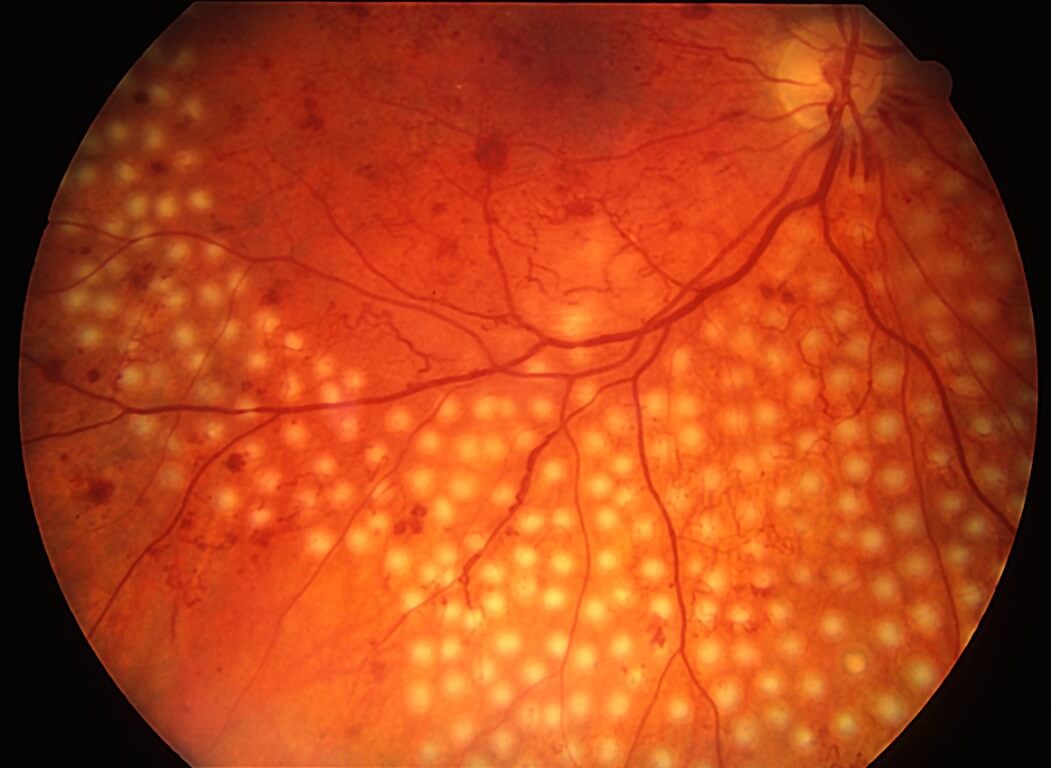
Retinal laser, also known as retinal laser therapy or retinal photocoagulation, is a medical procedure that uses focused laser light to treat various eye conditions affecting the retina and surrounding structures. Retinal laser therapy can be used in the following situations:
- Treatment of Retinal Diseases: Retinal laser therapy is a key treatment option for several retinal diseases and conditions, including diabetic retinopathy, retinal tears, retinal detachments, macular edema, and certain vascular disorders. It is particularly effective in managing these conditions when they involve abnormal blood vessels or retinal tissue.
- Sealing Leaking Blood Vessels: In diabetic retinopathy and some other retinal conditions, abnormal blood vessels can leak fluid and blood into the retina, causing vision impairment and potential vision loss. Retinal laser therapy can be used to seal these leaking vessels, preventing further damage and reducing the risk of vision deterioration.
- Retinal Tear and Detachment Repair: For retinal tears and detachments, retinal laser therapy is often employed to create small burns or adhesions around the tear or detachment site. This helps reattach the retina to its proper position and prevents the condition from worsening.
- Control of Abnormal Blood Vessel Growth: In cases of neovascularization (abnormal blood vessel growth) in the retina, such as in wet age-related macular degeneration or proliferative diabetic retinopathy, retinal laser therapy can be used to target and reduce these abnormal vessels. This helps stabilize the condition and prevents further vision loss.
- Macular Edema Management: Retinal laser therapy can help manage macular edema, a swelling of the macula (the central part of the retina responsible for detailed vision). Laser treatment can reduce the fluid buildup and improve central vision in conditions like diabetic macular edema.
- Minimally Invasive: Retinal laser therapy is considered a minimally invasive procedure. It typically does not require surgical incisions or general anesthesia, making it a more comfortable and accessible treatment option for many patients.
- Preservation of Vision: The primary goal of retinal laser therapy is to preserve and, in some cases, improve a patient’s vision. By targeting the underlying causes of retinal conditions, laser therapy can help prevent further vision loss and improve a patient’s overall visual function.
While retinal laser therapy can be highly effective, it may require multiple sessions for some conditions, and the results can vary depending on the severity of the disease and individual patient factors. It is typically performed by experienced retina specialists. Early diagnosis and appropriate treatment with retinal laser therapy can play a critical role in preserving and improving a patient’s vision, making it an essential tool in the field of ophthalmology.
